Image clarity is how well an imaging system can accurately reproduce the details and features of...
Image clarity is how well an imaging system can accurately reproduce the details and features of its scene. Clarity represents the overall quality and sharpness of an image. Surgeons need excellent image clarity in order to see small biological features in tissues and organs, which have depth and curves. Resolution, a key factor for image clarity, specifically describes the ability to distinguish between two features that are close together. Another factor for image clarity is depth of field, which we will explore in Image Clarity: Part 2.
The Challenge of Resolution
The point where two features can be distinguished is the system's resolution limit. Imagine trying to read increasingly smaller text on an eye chart. At some point, the letters start to blur together and become unreadable.
A similar “eye test” for fluorescence imaging systems can be achieved using a resolution target, which contains groups of three parallel lines that get progressively smaller. The system's ability to distinguish these bars determines its resolution. See the examples below showcasing high and poor resolution. (If they look the same to you, it might be time to visit your optometrists)
 |
 |
| In the high-resolution image, the individual lines are easily distinguished as three distinct stripes. | In the poor-resolution image, these same fine lines blur together into what looks like a single rectangle. |
The smallest group of lines where the stripes are distinct indicates the system's resolution limit. Scientists and engineers use a benchmark called the Rayleigh Criterion to determine when the imaging system can officially "resolve" two features as separate objects rather than seeing them as one blurred feature. This criterion defines the threshold as when contrast between two features reaches 26.4%.
Characterizing Your System's Resolution
To characterize, we can use a resolution target which traditionally has black and white bars that get progressively smaller. QUEL’s fluorescent resolution target replaces the black and white bars with fluorescence and background signals.
- Using your fluorescent imaging system, take images of a tissue-equivalent resolution target under typical conditions (working distance, ambient lighting, system settings).
- Identify groups of lines and elements to assess. For each peak in each element, calculate contrast C using the equation:
(Imax - Imin) / (Imax + Imin), where Imax is the maximum intensity and Imin is the minimum intensity. For each element, take an average. This can be done semi-automatically with the QUEL-QAL Python library. - Plot contrast vs. spatial frequency (line-pairs per mm). This can be done automatically with the QUEL-QAL Python library. Line-pairs per mm provide a metric for resolution, which can easily be converted to pixel resolution.
 |
 |
 |
| QUEL Imaging Resolution Target | Example false-cover fluorescence image QUEL Imaging Resolution Target. | A plot of contrast vs. image resolution generated using the QUEL-QAL library. |
For detailed step-by-step instructions, follow Use Guide: Fluorescence Resolution Targets.
Resolution: More Than Just Pixel Count
A common misconception is that resolution is simply about the number of pixels in your camera. While having a high-resolution sensor (like HD or 4K) is important, the actual spatial resolution of your system depends on multiple factors:
- Optical Resolution: The lens system often sets the fundamental limit for resolution, regardless of sensor quality. Even with a 4K camera, poor optics will blur fine details before they ever reach the sensor. This is just like wearing the wrong prescription glasses.
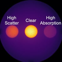
- Tissue Interactions: When imaging biological structures, light must travel through tissue where it scatters and spreads out. This scattering effect fundamentally limits how clearly we can see deeper structures, much like how fog makes distant objects appear less distinct. Simply adding more pixels or magnification cannot overcome this physical limitation.
- Digital Resolution: Modern cameras can detect subtle variations in light intensity through increased bit depth (from 8-bit to 16-bit). While this helps distinguish fine differences in brightness - like seeing more shades of gray - it doesn't necessarily help separate small features that are close together.
Best Practices for Development
Understanding and correctly measuring resolution is crucial for developing effective imaging systems. While the underlying physics can be complex, focusing on practical measurements and real-world performance will help ensure your system meets clinical needs:
- What is the expected feature size for your specific clinical indication? You do not need cellular resolution if you’re looking for blood vessels.
- What field of view does the surgeon need to visualize? Magnification, like reading glasses, can help, but with more magnification you lose context of the surrounding environment.
- Do you expect non-specific background fluorescence? Remember contrast between bright and dark helps define resolution.
- What form-factor imaging system do you require for your indication? The size and type of imaging system (e.g. hand-held, minimally invasive) put constraints on the optical design.
- What are your system characteristics? The tests described here can help characterize your ideal performance, and allow you to further optimize your system design.
Test your system using the same settings (camera exposure time, camera gain, working distance, ambient lighting conditions, etc.) that will be used in clinical practice. If your system has different operating modes, characterize each one separately. By understanding these principles and following proper testing procedures, you'll be better equipped to develop and validate your fluorescence imaging system's resolution.
For more detailed guidance on system characterization and standardization, refer to the AAPM TG311 guidelines. Implementation tools and reference targets are available to help you meet these standards effectively.
Interested in characterizing your imaging system or developing a customized fluorescence reference target? Contact QUEL Imaging!