

Look at the back of your hand. Beneath the surface, faint lines trace their way toward your wrist: cool-toned, blue-green, almost translucent.
The color is striking because it does not match what we know about blood.
Blood is red whether oxygen-rich or oxygen-poor, whether inside the body or outside it.And yet, those veins often appear blue - often with a green cast.
The mismatch is familiar, and it invites two closely related questions:
- Is venous blood somehow blue?
- If not, where does that color come from?
The answer is not in the blood itself. It emerges from an interplay of effects:
- how light enters the skin,
- how different wavelengths scatter and penetrate different depths,
- how blood absorbs part of that light,
- and how the visual system interprets the light reflected back to the eye.
The starting point is the optical behavior of skin.
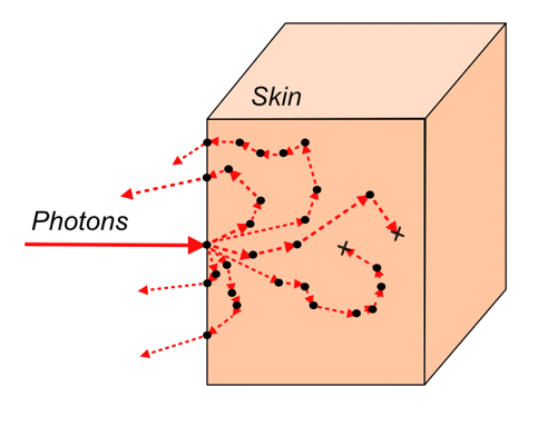 Skin is a turbid medium: a material in which light is both absorbed and strongly scattered.1 With visible light , scattering dominates. When light enters the skin, it does not travel straight down and back up; it is redirected repeatedly, diffusing through layers of tissue before eventually re-emerging.
This scattering depends on the color of light, or wavelength. Shorter wavelengths, toward the blue end of the spectrum, scatter more strongly and tend to remain near the surface. Longer wavelengths, toward the red, scatter less and can penetrate deeper before being redirected back out.
This difference in penetration depth is central. It means that the color of light returning from the skin depends not just on the surface, but on how deeply different wavelengths probe the tissue beneath.
Now consider a vein beneath the skin.
A vein is a volume of blood located some distance below the surface. When light enters the skin above it, different wavelengths interact with the vessel to different extents.
Blue light is largely confined to superficial layers and does not reach the vein in significant amounts. It is scattered back out with relatively little influence from the vessel.
Red light, by contrast, penetrates more deeply and is more likely to encounter the vein. Once there, it interacts with blood, which absorbs part of that light, particularly in the red and near-infrared regions, and more strongly for deoxygenated blood.
The result is a local difference in the reflected light: the region above the vein reflects less red light than the surrounding skin.
The key point is comparative. The difference is not primarily in how much blue light is reflected, but in how much red light is reduced relative to nearby tissue.
Much of our current understanding of this effect traces back to a detailed work by Kienle and colleagues in the 1990s.2 Using Monte Carlo models of light transport in tissue, they showed that a subsurface vein reduces long-wavelength (red) reflectance more strongly than short-wavelength (blue) reflectance, without ever making blue light dominant in absolute terms.
At first glance, this might suggest that the vein should simply appear slightly darker, with less red, rather than distinctly blue. The remaining step lies in how that difference is interpreted visually by our eyes and brain.
Human color perception depends not only on the light arriving from a given point, but also on how that light compares to its surroundings. The visual system continually evaluates these differences and assigns color based on relative contrast.
A key result follows from this: the light returning from above a vein can still contain more red than blue overall, and yet the vein is perceived as blue. The appearance depends on local relative contrast, not on absolute optical reflectance spectra.
When the region above a vein returns less red light than the surrounding skin, it creates a relative red deficiency. This imbalance shifts the perceived color by our eyes toward blue.
The same work also clarified the role of perception. Drawing on ideas closely related to Retinex theory,3,4 it showed that perceived color depends on ratios of reflected light across regions rather than isolated reflectance spectra. In that framework, the color of the vein is determined by how its reflected light compares to adjacent skin across different wavelength bands.
 These predictions have been confirmed experimentally. Spectral measurements, both in living tissue and in controlled models, show that reflectance above veins is reduced more strongly in the red portion of the spectrum than in the blue.
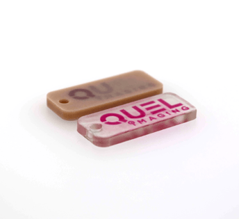
Recent work by Wagner et. al.5 has examined this under controlled conditions using tissue-mimicking phantoms - similarly to how we make phantoms at QUEL Imaging.
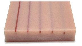
In these experiments, synthetic materials are engineered to match the optical properties of human skin, and cylindrical channels are embedded to represent blood vessels. These channels are filled with materials that mimic the absorption characteristics of oxygenated and deoxygenated blood. By varying depth, diameter, and oxygenation levels, researchers isolated how each factor contributes to appearance.
The results reinforce a consistent picture.
- There is a limited depth range (~1 mm) where veins are most visible and most likely to appear blue.
If a vessel is too shallow, shorter wavelengths can also reach it, and the color shifts toward darker or more purplish tones. If it is too deep, its influence on reflected light becomes too weak to produce a visible contrast. - Diameter also plays a role.
Larger vessels affect a broader region of the reflected light field and produce a stronger contrast with surrounding tissue. Very small vessels do not significantly alter the reflected signal over a large enough area to be perceived clearly. - Oxygenation contributes as well.
Deoxygenated blood absorbs more strongly in the red, which enhances the reduction in long-wavelength reflectance and makes veins more likely to appear blue than arteries under similar conditions.
Hyperspectral measurements in these systems make the mechanism explicit. At shorter wavelengths, reflectance above a vein changes only modestly. At longer wavelengths, particularly in the red, the reduction is more pronounced. The overall spectrum remains weighted toward red, but relative to surrounding tissue, it is depleted—producing the observed color shift.
It is also useful to clarify what is not responsible for this effect.
Veins are not blue because blood is blue. Blood remains red under all physiological conditions.
The effect is not simply a matter of the skin acting as a color filter.
And while scattering is essential, this is not a straightforward example of Rayleigh or Tyndall scattering.
The appearance depends on the combined roles of scattering, It arises from wavelength-dependent scattering, hemoglobin absorption, and visual perception of color differences.
Light absorption, tissue geometry, and visual perception.
Taken together, the explanation is straightforward:
- Light enters the skin and is scattered, with shorter wavelengths remaining near the surface and longer wavelengths penetrating more deeply.
- Red light reaches subsurface veins more effectively than blue light.
- Blood absorbs part of that red light, reducing the amount that returns.
- The region above the vein therefore reflects less red light than the surrounding skin.
- The visual system interprets this relative reduction as a shift toward blue.
The question “if blood is red, why are veins blue?” assumes that color is a direct property of an object. In this case, it is not.
The color of a vein is the result of light moving through tissue, interacting with structure and composition, and being interpreted by the visual system. It is not the color of blood alone, but the outcome of how that blood is seen.
What begins as a mismatch turns out to be a matter of perspective. Blood is red. Veins appear blue-green under the skin. The discrepancy vanishes once the physics of light in tissue and the way our visual system interprets color accounted for.
QUEL Imaging helps teams understand and translate optical technologies into consumer and clinical products with optically-tuned tissue phantoms. Learn more about our multispectral tissue phantom capabilities here or reach out to learn how we can help your team accelerate device development.
References:
1. W. F. Cheong, S. A. Prahl, and A. J. Welch, “A review of the optical properties of biological tissues,” IEEE J. Quantum Electron. 26(12), 2166–2185 (1990) [doi:10.1109/3.64354]. https://ieeexplore.ieee.org/document/64354
2. A. Kienle et al., “Why do veins appear blue? A new look at an old question,” Appl. Opt. 35(7), 1151 (1996) [doi:10.1364/AO.35.001151]. https://pubmed.ncbi.nlm.nih.gov/21085227/
3. E. H. Land and J. J. McCann, “Lightness and Retinex Theory,” J. Opt. Soc. Am., JOSA 61(1), 1–11, Optica Publishing Group (1971) [doi:10.1364/JOSA.61.000001]. https://opg.optica.org/josa/abstract.cfm?uri=josa-61-1-1
4. J. J. McCann, “Retinex at 50: color theory and spatial algorithms, a review,” JEI 26(3), 031204, SPIE (2017) [doi:10.1117/1.JEI.26.3.031204].
https://www.spiedigitallibrary.org/journals/journal-of-electronic-imaging/volume-26/issue-3/031204/Retinex-at-50--color-theory-and-spatial-algorithms-a/10.1117/1.JEI.26.3.031204.full
5. M. Wagner et al., “Visual appearance of blood vessels: a phantom study,” Biomed. Opt. Express 17(2), 1049 (2026) [doi:10.1364/BOE.579246]. https://opg.optica.org/boe/fulltext.cfm?uri=boe-17-2-1049