Fluorescence imaging systems face a fundamental challenge: detecting signals through tissue, which...
Key Tools for Characterizing Fluorescence Imaging Systems
A Look at Optical Reference Targets
For developers and users of fluorescence imaging systems, ensuring optimal system performance is essential. One of the most effective ways to do this is by using optical reference targets—specialized phantoms designed to evaluate specific performance metrics. These tools help quantify system sensitivity, depth detection, and resolution, providing reliable, standardized testing methods that mimic real-world surgical conditions.
Here’s a closer look at three main types of reference targets which follow international consensus recommendations (AAPM TG311), and how they’re used in fluorescence imaging system characterization.
1. Concentration Sensitivity Target
Detecting a fluorescence signal accurately at varying concentration levels is critical for system performance. In the past, this was done with serial dilutions of liquid fluorophore solutions, but these were time-consuming, difficult to reproduce, and could contaminate the imaging system.
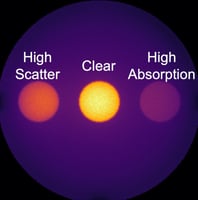
The Concentration Sensitivity Target addresses these limitations. It is a stable, solid phantom containing wells with a range of fluorophore concentrations, along with scattering and absorption properties that closely resemble tissue. This allows for straightforward testing and reliable quantitative analysis of three key metrics:
- Noise Floor: The lowest concentration at which fluorescence can be distinguished from ambient background light.
- Saturation Point: The concentration level at which the system can no longer quantify higher concentrations.
- Linearity Range: The concentration range within which the system can perform quantitative analysis accurately.
2. Depth Sensitivity Target
Understanding how deeply an imaging system can detect fluorescence is vital, especially for procedures requiring visualization beneath tissue layers. The Depth Sensitivity Target provides a way to test this.
This target uses a single concentration of fluorophore with non-fluorescent material layers stacked on top, with each well having a different thickness. By analyzing the fluorescence intensity across these wells, the system’s depth detection limits are quantified. This test simulates real tissue conditions and helps developers assess how effectively the system can detect fluorescence through varying tissue depths.
3. Resolution Target
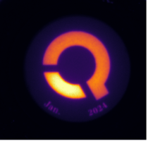
Resolution is essential for identifying fine details and distinguishing between close features in fluorescence imaging. The Resolution Target uses a standard test pattern etched on glass placed over a fluorescent background.
This pattern includes progressively smaller groups of three-line elements, allowing the system’s resolution to be assessed by measuring contrast at each element. This test determines the smallest feature size that the system can reliably detect, helping developers understand the system’s limits in terms of detail and clarity.
Why Optical Reference Targets Matter
These three optical reference targets—concentration sensitivity, depth sensitivity, and resolution—offer an efficient and reliable way to test imaging systems. By providing controlled, repeatable testing conditions, they allow both system developers and users to obtain accurate metrics on system performance, ensuring these imaging systems work effectively in clinical settings.
Key Takeaways on Using Reference Targets
- Consistency and Reliability: Reference targets offer a stable, reproducible way to test system performance, avoiding the variability of liquid-based methods.
- Comprehensive Performance Metrics: Each target focuses on a key aspect—concentration, depth, and resolution—providing a holistic view of system capabilities.
- Realistic Testing Conditions: These phantoms mimic the optical properties of tissue, giving developers insight into how systems will perform in surgical environments.
Optical reference targets are indispensable tools in fluorescence imaging system development and quality assurance. By quantifying essential performance metrics, they enable more accurate, consistent imaging results, supporting better surgical outcomes and advancing fluorescence-guided imaging technology.


