A Look at Optical Reference Targets For developers and users of fluorescence imaging systems,...
Tissue Optical Phantoms: Essential Tools for Advancing FGS
Fluorescence-guided surgery (FGS) gives surgeons enhanced precision by illuminating specific tissues during procedures, but developing these systems requires thorough testing to ensure reliable performance under real surgical conditions. Tissue optical phantoms—engineered materials that mimic human tissue properties—are essential in this process, offering a controlled and reproducible way to assess and optimize FGS systems.
Challenges in Testing FGS Systems
FGS imaging systems go through rigorous testing and regulatory review to ensure they perform effectively. Traditional characterization methods often involve serial dilutions of contrast agents like ICG to gauge sensitivity, but these liquid dilutions rarely represent real-world performance, where biological tissues and blood can impact sensitivity.
In practice, some developers resort to improvised methods, such as placing contrast agents under bacon or raw chicken to replicate tissue, but these lack reproducibility and may not be representative of human tissue. Phantoms solve this issue by providing standardized testing tools that reflect the optical properties of human tissue in a consistent, controlled way.
How Phantoms Mimic Tissue
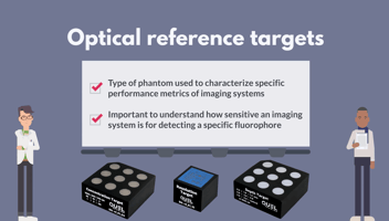
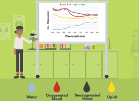
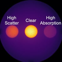
Phantoms are designed to replicate specific tissue characteristics, particularly light scattering and absorption. By carefully combining materials that mimic these optical properties, phantoms simulate tissues like muscle, fat, or tumor. When paired with contrast agents, they provide realistic performance feedback, helping refine system sensitivity, contrast, and image quality—crucial for effective intraoperative imaging.
Applications in Development and Quality Assurance
In early development, phantoms enable engineers to adjust FGS device settings under conditions that closely mirror those in surgery. Phantoms are also valuable in regulatory review, as they produce reliable performance data in a repeatable environment.
Beyond development, phantoms are increasingly used in quality assurance to ensure devices continue to meet performance standards over time. When comparing the performance of different systems, such as during regulatory submissions or multi-center clinical trials, phantoms can serve as standardized references to provide consistent, qualitative metrics.
Key Takeaways: The Value of Phantoms in FGS
- Standardization: Phantoms offer a consistent testing environment, overcoming the variability of ad hoc methods and ensuring reliable, reproducible data.
- Realism: By accurately mimicking tissue properties, phantoms provide a realistic benchmark, helping developers understand how an FGS system will perform in surgery.
- Regulatory Support: Phantoms produce data in a reproducible manner that can help meet regulatory standards, which is essential for system approval and compliance.
- Quality Assurance: Phantoms are essential for ongoing device testing and for comparing data across different imaging systems or multiple testing sites.
Tissue optical phantoms are indispensable in developing, validating, and maintaining high-quality FGS imaging systems. By providing a realistic and reproducible testing environment, phantoms enable more effective, consistent system performance, ultimately supporting better outcomes in fluorescence-guided surgery. See more of what QUEL Imaging has to offer to address your FGS needs.


