A preprint providing guidelines for performance evaluations of fluorescence-guided surgery (FGS)...
Optical Surgical Navigation: WMIC 2022
The World Molecular Imaging Congress was hosted in Miami at the end of September. Each year at this conference, the World Molecular Imaging Society's Optical Surgical Navigation (OSN) interest group hosts a workshop to highlight the work being done in the field to address the OSN mission to "advance and support optical molecular imaging education, innovation, standardization, and translation to clinical use." This workshop was moderated by Jim Delikatny, PhD (University of Pennsylvania), Summer Gibbs, PhD (Oregon Health & Science University) and Stephan Rogalla, MD PhD (Stanford Medicine).
This workshop brought together the field's leading chemists, surgeons and engineers to discuss the current state of development and what is needed to advance the future of intraoperative molecular imaging. The meeting started with Philip Low, PhD (Purdue University) discussing his experience as a chemist developing tumor-targeted fluorescence dyes. It was fascinating to hear how Dr Low's team advanced the development of what is now known as OTL38, or Cytalux, starting with animal studies in 2002. This led to early in-human studies beginning in 2008, and after numerous trials the recent FDA approval for use in adults with ovarian cancer to, "enhance the ability of surgeons to identify deadly ovarian tumors that may otherwise go undetected".
One of the unexpected challenges during clinical studies was identifying interested surgeons and medical centers equipped with a suitable imaging system. Once appropriate sites were identified, implementing protocols for interpreting visualization data and defining outcomes, was the next key step. Many of the current ICG-based imaging systems can visualize OTL38, but the imaging techniques are slightly different. ICG is primarily intended for vascularity or flow assessment, so small imaging artifacts are easily ignored. However, with targeted fluorescence, small artifacts may be more difficult to distinguish from small cancerous lesions, leading to a higher rate of false positives.
Additional presentations were given by surgeons working on clinical studies using a variety of targeted and non-targeted fluorophores, primarily for tumor resections. While many investigations have demonstrated targeted fluorophores for enhancing intraoperative surgical guidance, intra-operative back-table imaging is a growing trend where resected sections are imaged and fluorescence is used to guide biopsy selection for pathology margin assessments. Ultimately, as has been stated by many surgeons, the only sure cure for cancer is to cut is all out, and fluorescence is a great tool to enhance a surgeon's vision.
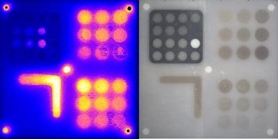
In an effort to standardize the field, Vasilis Ntziachristos, PhD presented on his group's efforts to develop phantoms and tools. Much of the pre-commercial work developed by the QUEL Imaging co-founders was inspired by this group, and is documented in a joint publication in the Journal of Biomedical Optics. Dr. Ntziachristos' main argument was that fluorescence can be easily imaged, but how accurate is the image? This is something we, and others in the field, continue to work on.
Finally, outside of the OSN working group meeting, it was very encouraging to see how some of our customers are using our products. Hans Ingleberts gave a presentation about the fluorescence lifetime imaging system his group is developing at Vrije University in Brussels. They have been using our targets during system development, and presented a demonstration video using our vessel phantoms.
As always, we're open to discuss your applications and how we can help. Have a question? Reach out and setup a call!