In the world of fluorescence imaging, both excitation intensity and emission detection sensitivity...
Precision Surgery, Fluorescence, and the Road to Real-World Adoption
A recap from the “Precision Surgery Intraoperative Molecular Imaging: 4th Clinical Trials Update” at UPenn
Last week, Alberto and I had the chance to attend the Precision Surgery Intraoperative Molecular Imaging: 4th Clinical Trials Update symposium at the University of Pennsylvania. It was a dense, energizing day that reinforced a simple but important truth:
The science behind intraoperative molecular imaging is no longer speculative. It’s here, it’s working, and it’s growing. But the challenge now is scaling adoption — through reimbursement, training, and culture change.
The meeting brought together surgeons, engineers, industry partners, and scientists to focus on one thing: how to move fluorescence-guided surgery and intraoperative molecular imaging from specialized centers into routine clinical practice.
From “To Cut Is to Cure” to Precision Surgery

The day opened with historical and clinical context from several surgical leaders at Penn.
Surgery is getting harder, not easier
Thoracic surgeon and conference organizer, Sunil Singhal, MD, opened the meeting by tracing the arc of cancer surgery:
The first reports of cancer surgery date back to 50 AD in Alexandria, Egypt, where cautery was used to remove a breast lesion. From 1834 the Pennsylvania Hospital Surgical Amphitheatre was used to demonstrate surgeries to up to 130 people at a time. Keep in mind, anesthesia was not demonstrated until 1846, so surgeries were a always a race. And sterile techniques were not mandatory in America until the early 1900s. The next major progression was in the early 1970s with the first commercially available CT imaging system, which expanded in 2001 to include PET/CT for assessing cancer progression. A few years later, in 2005 the first fluorescence guided imaging system received FDA clearance to assess tissue perfusion with ICG.
Today, about 1 million cancer surgeries are performed each year in the US (5 million worldwide). The goal of every one of those cases is simple in theory: cure the cancer. But, as Singhal emphasized, roughly 40% of patients recur—half locally, half systemically. At the same time, surgery is getting more complex:
-
Radiology finds smaller and smaller lesions
-
Robotic platforms have improved access but removed the surgeon’s ability to palpate
-
Frozen section pathology is useful, but not always enough to ensure negative margins in real time
The old mantra, “To cut is to cure,” is no longer sufficient. As Singhal put it:
“To cut well is to cure. To cut poorly is to harm.”
That’s the space where intraoperative molecular imaging fits: helping surgeons cut well. The answer is increasingly tied to how well we can see tumor and critical structures during the operation.
The Science Is There: Probes, Trials, and Real Clinical Endpoints
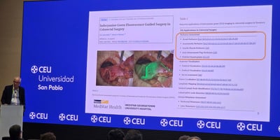
One of the clearest messages from the day: we are not waiting for hypothetical tools. Many of these agents and devices are in, or past, Phase II and Phase III trials, with clear clinically significant events (CSEs). The table below provides a brief overview of some fluorophores that were discussed at the meeting.
Fluorophore |
Generic Name / Description |
Indication |
QUEL Equivalent Product Line |
| ICG | Indocyanine Green | Mainly perfusion, lymphatic networks | ICG |
| ALA-PpIX | 5-Aminolevulinic acid → Protoporphyrin IX | Glioma surgery, also used in skin pre-cancer and PDT treatments | Contact Us! |
| OTL38 | Pafolacianine (folate-receptor targeted dye) | Ovarian cancer and Lung Cancer visualization | Q800 |
| LUM015 | Pegulicianine (cathepsin-activated Cy5 fluorescent probe) | Breast cancer margin assessment, Trials in colorectal cancers | Contact Us! |
| SGM-101 | CEA-targeted antibody-dye conjugate | Trials in colorectal cancer visualization | Q700 |
| VGT-309 | Abenacianine (cathepsin-targeted NIR imaging agent) | Trials in lung cancer visualization | ICG |
| FG001 | uPAR-targeted ICG | Trials in glioma visualization, early-phase trials on head and neck cancer | ICG |
| LGW16-03 | nerve-specific red-fluorescent small molecule | Preclinical trials for nerve sparing surgeries | Contact Us! |
| ALM-488 | Bevonescein (nerve-illumination peptide-dye) | Head and neck nerve imaging | Not available |
| JAS239 | NIR-fluorescent choline kinase α inhibitor | Preclinical trials in lung cancer visualization | Contact Us! |

This is not an exhaustive list, and there was a clear trend to develop contrast mechanisms that improve sensitivity and specificity of cancer detection. While ICG is a non-specific dye, when used it certain regimens it can localize in tumors highlight sentinel lymph nodes. Molecular targeting provides higher specificity, and activatable probes using cathepsins or pH triggers can further reduce non-specific background. But even with these amazing advancement in probe development, there are still many challenges for clinical adoption.
The Bottlenecks: Reimbursement, Workflow, and Training
If the science is compelling, why isn’t every OR using fluorescence? A big part of the answer came in the form of very practical, sometimes blunt, comments from the speakers.
Reimbursement and RVUs
Surgeons live in a world of RVUs and CPT codes. As Singhal pointed out:
-
Industry and VC care about reimbursement
-
Surgeons care about whether a technique adds value and whether it’s recognized and compensated in their workflow
Examples like CPT codes 77001 (fluoroscopy) and 76998 (ultrasound) were cited as models. For fluorescence to be widely adopted, we’ll likely need:
-
A clear CPT framework for fluorescence imaging
-
The ability to bill for interpretation and decision-making, not just dye injection
-
Real RVUs that reflect the added time, complexity, and value of improved care
Without that, fluorescence risks being treated as a “nice add-on,” rather than a supported standard of care.
Workflow and device fragmentation
Another recurring theme: even when an agent is approved and reimbursed, the hardware ecosystem is messy.
-
Every specialty seems to have its own camera form factor
-
Open systems, laparoscopic towers, robotic platforms, loupes, endoscopes—each has different capabilities
-
Overlay, co-registration, and real-time interpretation are not yet standardized
This fragmentation makes it harder for teams to adopt a technique and trust it in high-stakes cases. And it makes it difficult for hospital administrators to compare options and identify the best system for a given set of indications.
Training the surgical team (not just the surgeon)
One of the most important points of the day was about education. Most surgeons don’t encounter fluorescence imaging in any serious way until they are already in practice. You can teach an “old dog” new tricks—but it’s much easier if exposure and training begin earlier:
-
In medical school
-
During residency and fellowship
-
In simulation labs, not just in live OR cases
A parallel was drawn to pilots and flight simulators: the technology can work perfectly, but without training, confidence, and pattern recognition, people will not use it correctly—or at all. Surgeons build habits over years, and advancement may not come from the most experience surgeons, so earlier training should accelerate adoption. The need for culture and generational change was tied back to the Max Planck quote:
“Science advances one funeral at a time.”
Growing the Field: From Niche to Norm
For me, the symposium underscored three big takeaways about the future of intraoperative molecular imaging:
-
The field is maturing. We have multiple Phase II and Phase III trials, FDA-approved agents, and clear evidence of clinically significant events: additional lesions, improved margin assessment, better staging.
-
The path to broader adoption is non-technical. The challenges now are:
-
Reimbursement structures and RVU alignment
-
Device and workflow standardization
-
Systematic training for surgeons, trainees, and the entire OR team
-
-
Early education is critical. If we want fluorescence imaging and molecular guidance to be routine, not exotic, they must show up early in the educational journey—in anatomy labs, simulation, and residency curricula.
Most people in that room were convinced of the potential. The next step is making sure the rest of the world can see what they see: that fluorescence is not just a cool visualization trick, but a tool that can shift outcomes, reduce complications, and make oncologic surgery more precise and humane.
Overall, we enjoyed our short visit to Philadelphia. It was great to catch up with so many customers and collaborators, hear the latest updates and discuss how we can help accelerate the translation of these amazing technologies. Let's keep the conversations going, reach out now!


