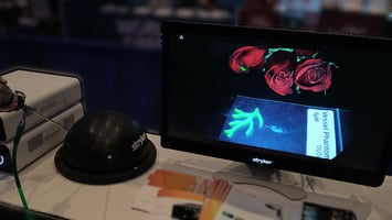
Fluorescence-guided surgery (FGS) gives surgeons enhanced precision by illuminating specific...
Fluorescence-Guided Surgery: Enhancing Intraoperative Visualization
Over the next few months, we will provide a series of educational posts discussing broad concepts and more specific applications of our targets and tools. If you would like to learn more, please check out our resources or reach out to us!
What is FGS?
Fluorescence-guided surgery (FGS) is an innovative technique designed to improve intraoperative visualization by using light beyond the visible spectrum. Although surgeons are trained to rely on their visual and tactile senses, FGS offers a powerful adjunct to enhance the identification of critical structures that may not be readily visible.
Mechanism of Fluorescence Imaging
The human eye is limited to detecting wavelengths within the visible spectrum (violet to red). FGS systems, however, employ specialized cameras and filters to detect specific wavelengths outside of this range. This is analogous to tuning a radio to a specific frequency—filters isolate precise wavelengths, allowing only those relevant signals to pass through to the camera sensor.
Functionality of FGS Systems
FGS systems generate two complementary images: one representing the standard visual field and another that highlights the fluorescence emitted by contrast agents. These agents absorb excitation light and emit fluorescence at different wavelengths, captured by the camera’s filters. This allows for real-time, enhanced visualization of the surgical field, giving surgeons the ability to toggle between conventional and fluorescence-enhanced views.
Interpreting Fluorescence Signals
Fluorescence images are initially captured in grayscale and are often displayed using colormaps applied by system manufacturers. These colormaps can modify the perceived intensity and contrast of the fluorescence signal, even when the underlying data remains unchanged. Understanding these variations is crucial for accurately interpreting the enhanced view.
Factors Influencing Fluorescence Detection
Several variables can affect the reliability and clarity of fluorescence signals:
- Proximity: The camera’s distance from the surgical site influences fluorescence intensity; moving closer increases the signal, while moving further away reduces it.
- Tissue Obstruction: Structures like adipose tissue can attenuate or obscure the fluorescence signal, potentially affecting visualization.
- Ambient Light Interference: The presence of ambient light sources emitting wavelengths similar to those of the fluorescence signal can lead to interference. This is comparable to the overlap of radio frequencies, where multiple signals are detected simultaneously, complicating interpretation.
Optimizing Fluorescence-Guided Surgery
While FGS significantly enhances contrast and aids in the differentiation of tissues, it is not without limitations. System manufacturers are continually refining technologies to improve both the sensitivity and specificity of these devices. As these advancements unfold, FGS will continue to evolve, providing increasingly precise and reliable imaging options for surgical applications.


