
The FDA recently released draft guidance for optical imaging drugs, including an appendix dedicated...
Bridging the Gap in Fluorescence-Guided Surgery: Insights from Lumicell
At the 2026 Stanford Symposium on Optical Surgical Navigation, Brian Schlossberg, VP of Innovation at Lumicell, shared a detailed and candid overview of the development and commercialization of their fluorescence-guided surgery platform. This talk highlighted not only the path to approval, but the broader structural challenges that can limit adoption if companies do not plan ahead.
The Clinical Need: Margin Assessment in Breast Surgery
One of the most important unmet needs in surgical oncology is intraoperative margin assessment during breast-conserving surgery. Incomplete resections often lead to follow-up procedures, which increases both patient burden and healthcare costs.
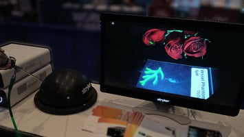
Lumicell’s approach, described as “Inject, Detect, Resect,” is designed to address this gap. Their system (LumiSystem™) combines:
- LUMISIGHT™ (pegulicianine), a fluorescent imaging agent
- Lumicell™ Direct Visualization System (DVS), an intraoperative imaging platform
LUMISIGHT™ is administered intravenously 2 to 6 hours prior to imaging. This allows surgeons to visualize residual cancer directly within the surgical cavity. The goal is straightforward: detect and remove residual disease in a single procedure.
A Decade-Long Development Path
The development timeline for Lumicell’s platform highlights the complexity of drug-device combinations:
- 2012: IND approval for their activatable fluorescent imaging agent LUMISIGHT
- 2012–2014: Phase I trials (N=15)
- 2015–2020: IDE feasibility studies across multiple phases and centers
- 2020–2022: Pivotal trial (N=406)
- March/April 2023: NDA (imaging agent) and PMA (device) submissions
- March 2024: FDA advisory panel vote (16-2-1 in favor)
- April 2024: Approval
Beyond the timeline itself, several key lessons emerged. A key takeaway from Dr. Schlossberg’s discussion was the importance of early and sustained engagement with the FDA. Aligning on endpoints that reflect both diagnostic performance and clinical utility is critical.
Most of the regulatory burden was driven by the imaging agent component, even though the system is used by the surgeon as a device. This dual-path approval, with CDER for the imaging agent and CDRH for the device, adds complexity that many developers underestimate.
Designing for the Indication
A central lesson from Lumicell’s experience was the importance of defining the indication early and designing everything around it.
For Lumicell, this meant aligning clinical trials, system design, and workflow integration around a specific surgical use case. Their primary endpoints included:
- Residual cancer detection (7.6% of patients)
- Sensitivity (49%)
- Specificity (86.5%)
At first glance, the sensitivity may seem low. However, performance must be evaluated against the current standard of care. The surgeon images the lumpectomy cavity after they’ve completed their standard of care procedure and done their best to remove the disease. A sensitivity of 49% means roughly half of the residual cancer that would otherwise have been left behind is detected in real time, in approximately seven minutes of intraoperative imaging — disease that otherwise might only have been identified at a second surgery or not at all. In this setting, even incremental improvements in resection completeness can have meaningful clinical impact.
Workflow and Training Matter
Successful deployment depends not just on the technology, but on operational execution. It requires:
- Coordinating imaging agent preparation and timing with the pharmacy
- Training surgeons, nurses, surgical and sterile processing staff
- Providing field engineering support for installation, setup and case support
The system is not inherently difficult for surgeons to use. The challenge is ensuring everything is ready when the surgeon needs it. Operational readiness plays a major role in adoption.
Reimbursement Strategy: Coordinating Coding, Coverage, and Payment
Drug-device combination technologies require coordinated reimbursement for both hospitals and providers. Hospitals need appropriate payment pathways for the imaging agent and associated facility costs, and providers must integrate the technology within surgical workflow and operative resource constraints. Sustainable adoption depends on reimbursement pathways that fit the technology into existing surgical workflows — clinical evidence alone does not drive uptake when the economic structure does not support it.
Lumicell recognized early that successful commercialization would require alignment across several reimbursement mechanisms and therefore pursued multiple reimbursement pathways concurrently with the clinical and regulatory program.
- Category III CPT add-on code for intraoperative fluorescence imaging during lumpectomy was approved by the AMA CPT Editorial Panel in 2024 and became effective Jan. 1, 2025. The code established a procedural reporting and payment pathway associated with physician use of the technology and represented an important milestone in the company’s reimbursement strategy.
- Centers for Medicare & Medicaid Services (CMS) HCPCS code for LUMISIGHT became effective January 1, 2025; establishing a standardized billing and reimbursement pathway for the imaging agent in the hospital outpatient setting.
- Transitional pass-through (TPT) status was granted by the CMS effective October 1, 2024; enabling separate Medicare payment at ASP+6% and establishing a clear reimbursement pathway for hospital outpatient adoption.
This strategy highlights a broader issue: U.S. reimbursement is built for therapies used repeatedly over time, with defined payment pathways. Intraoperative fluorescence imaging is different — a single-use agent used once during surgery to inform decisions in real time. No existing category was designed for that shape, so commercialization requires building coding, coverage, payment, and provider economics in parallel with the clinical and regulatory program. The path is engineered, not accessed.
The Disconnect Between Imaging Agent and Device Development
Lumicell’s approach stands out because they developed the imaging agent, device, and software as a unified system.
In many cases, there is a disconnect between:
- Imaging agent developers, who follow development pathways
- Imaging system developers, who operate within device frameworks
These groups are often developed and operated separately and are regulated by different parts of the FDA on different timelines. All of this creates friction. Lumicell’s integrated approach stands in contrast to this typical fragmentation.
A device company cannot wait for an imaging agent to be approved before starting development. This forces early partnerships or, as in the case of Lumicell, pushes imaging agent developers to build their own systems. Both approaches carry different types of risk.
Draft FDA guidance on combination products has started to address these challenges. Industry feedback, including input from QUEL Imaging, has emphasized the need for more coordinated pathways. For now, developers still have to navigate parallel processes that are not fully aligned.
Strategic Tradeoffs in System Design
A key constraint in development is the need to lock the device design early. Once clinical trials begin, changes to the system, especially software, can trigger additional regulatory requirements.
This creates a tradeoff between:
- Continuing to improve the system
- Maintaining a stable configuration for clinical validation
It also raises a broader question. As more general-purpose imaging systems become available, will future imaging agents rely on existing platforms, or will custom systems remain necessary for optimal performance?
Looking Ahead
Lumicell’s journey highlights both the promise and the structural complexity of fluorescence-guided surgery. Their success demonstrates that an integrated approach can work. At the same time, it highlights the structural challenges that still exist.
For the field to grow, better alignment is needed across:
- Regulatory pathways
- Reimbursement models
- Development timelines
At QUEL Imaging, we see this as a key opportunity. Bridging the gap between imaging agent developers and imaging system designers will be essential to advancing the field and improving surgical outcomes. If you’re navigating similar challenges in fluorescence-guided surgery, we’d be glad to connect. At QUEL Imaging, we work with teams across imaging agent and device development to support validation, training, and clinical translation.
Important Safety Information
What is LUMISIGHT (pegulicianine) and Lumicell DVS?
- LUMISIGHT (pegulicianine) is an optical imaging agent and Lumicell DVS is a fluorescence imaging device. LUMISIGHT and Lumicell DVS, in combination, are used in adults with breast cancer to help detect any remaining cancerous tissue at the surgical site following removal of the primary specimen during a lumpectomy procedure.
What is the most important information to know about LUMISIGHT?
- Hypersensitivity Reactions: LUMISIGHT may cause serious hypersensitivity reactions, including anaphylaxis. Anaphylaxis has occurred in 4/726 (0.6%) of patients in clinical studies. Tell your doctor if you have any history of hypersensitivity reactions to pegulicianine or to contrast media or products containing polyethylene glycol (PEG). Your healthcare provider should have emergency resuscitation drugs, equipment, and trained personnel available during the use of LUMISIGHT. Healthcare providers should monitor all patients for hypersensitivity reactions and if one is suspected, immediately discontinue the injection and initiate appropriate therapy.
- The most common side effects (≥1%) include hypersensitivity and an abnormal color in urine.
What additional important information should I know about LUMISIGHT and Lumicell DVS?
- Adjunctive Use: Lumicell DVS is for use as part of the lumpectomy procedure and is not a replacement for the standard of care lumpectomy procedure and pathology.
- Risk of Misdiagnosis: The absence of a signal in surgery does not rule out cancer. Additionally, a positive signal may be seen in noncancerous tissue.
- Interference from Dyes Used for Sentinel Lymph Node Mapping: Your healthcare provider should avoid use of dyes before imaging the lumpectomy cavity in patients who have received LUMISIGHT.
- Other Risks: Using the Lumicell DVS handheld probe may cause tissue damage or infection.
You are encouraged to report negative side effects of prescription drugs to the FDA. Visit MedWatch or call 1-800-FDA-1088. Please see the LUMISIGHT Prescribing Information, including Boxed Warning, and Lumicell DVS Instructions for Use. For complete product information www.LumiSystem.com.