At the 2026 Stanford Symposium on Optical Surgical Navigation, two consecutive presentations...
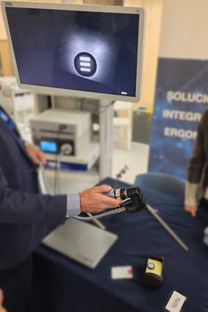
In November, I joined surgeons, engineers, and industry partners in Madrid for the ISFGS European Chapter Meeting. The tone of this meeting contrasted with the clinical trial updates presented the previous week at the Precision Surgery Intraoperative Molecular Imaging symposium. Instead of focusing on trials and future developments, the discussions in Madrid highlighted how fluorescence is being used in operating rooms today.
 Session after session demonstrated the versatility of indocyanine green (ICG) in enhancing a surgeon’s vision across a wide range of indications. These included perfusion assessment, lymphatic mapping, and several emerging applications. The talks showed that clinical teams are moving beyond treating fluorescence as an experimental tool and are increasingly integrating it into routine workflows to improve the standard of care.
1. From “Does It Work?” to “Why Didn’t You Use It?”
In colorectal, liver, and breast surgery in particular, fluorescence is no longer framed as experimental. For many speakers, ICG has become part of their default toolkit for:
- Assessing anastomotic perfusion
- Guide liver resections and segmental anatomy
- Mapping lymphatics and sentinel nodes
2. Workflow and Culture Are the Bottlenecks
The technology is here: approved agents, capable imaging systems, and growing evidence. The limiting factors are:
- How teams organize their workflow around fluorescence
- How early and how well clinicians are trained
- How comfortable surgeons feel trusting a new visual modality in high-stakes decisions
Speakers repeatedly emphasized the need to train entire teams, not just surgeons, and to embed fluorescence into everyday practice rather than treating it as a special add-on. There was also a discussion of training the next generation of surgeons, who are less stuck in their current habits.
3. Quantification Is Coming
Across vascular, colorectal, hepatic, pancreatic, billiary and limb salvage talks, there was strong momentum toward quantitative fluorescence:
- Moving from “it looks bright enough” to defining thresholds
- In-flow and out-flow rates are promising metrics
- Standardizing camera distance, gain, exposure, and timing
- Linking signal metrics to hard outcomes like leaks, wound healing, and graft viability
The consensus: we need robust, validated quantification tools that translate signal intensity into actionable guidance.
4. Expanding Applications and Creative “On-the-Ground” Solutions
Fluorescence is expanding into:
- Metabolic and bariatric surgery
- IBD and inflammatory diseases
- Gynecologic and urologic procedures
- Vascular surgery and diabetic foot care
- Neonatal and even veterinary applications
5. Beyond Green: A Multimodal, Multispectral Future
Looking ahead, several talks pointed to a future where ICG is just one piece of a broader imaging ecosystem:
- Tumor-targeted fluorophores are the next generation
- Multispectral and hyperspectral imaging
Our Role: Training, Standardization, and Support
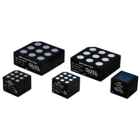
For us, this meeting reinforced that standardized, realistic training tools—like the Mobula-IGM SLNB phantom—play an important role in the next phase of adoption. It’s not enough to ship a camera or a contrast agent; teams need:
- Reproducible, anatomy-relevant models
- Clear protocols for dose, timing, and imaging
- Opportunities to build team-level muscle memory before stepping into a live case.
We left Madrid both energized and humbled. The clinical community is pushing fluorescence-guided surgery forward at remarkable speed, and we’re grateful to contribute a small part by supporting training, standardization, and optical characterization.
If you’re interested in ICG-based fluorescence for SLNB in breast cancer or in training tools for your OR team, we’d be happy to continue the conversation. We help teams develop training tools and quantification methods for fluroescence guided surgery all the time. Reach out to see how we can work together and move the field forward.